Cobra outputs
The CoBra project aims to improve the quality of both diagnosis and treatment of localised cancers, by developing a new medical robot for adaptive brachytherapy (BT) and biopsy under Real-time MRI guidance.
Capitalizing on robot precision and MR imaging quality with RT feedback, the objectives are to improve precision of the tumor tissue sampling (biopsy module) as well as the placement of the radioactive seeds with optimized dose planning while minimizing the tissue damage (nerves and blood bundles) thanks to the optimized trajectory calculation and an active steerable needle. All those features are integrated within the MRI compatible robot guide completing the biopsy and BT module and allowing a fast RT MRI guided intervention.
In parallel, a bioinspired and active Prostate Phantom reproducing the MRI environment was designed for robot validation and training purpose.
OUTPUTS
BIOPSY MODULE – LOCALIZED CANCERS DIAGNOSIS AS PART OF INTEGRATED COBRA ROBOTIC SOLUTION
Demcon Mechatronics, Delft, the Netherlands
The MRI Safe biopsy system has been developed within the CoBra Project as part of the integrated robot serving the diagnosis of soft tissues for detection of localized tumors. The creation of the Biopsy system required selection of MR-compatible components like actuators, sensors as well as materials. Additionally, a lot of effort has been put into improvement of the biopsy quality through precision and correct speed appliance when firing the sampling needle. The mechanical design has been optimized and adapted to fit into the MRI Scanner.
RT DOSE CALCULATION
Current treatment planning software include both the use of MRI and CT for dose calculation. CT is still needed for an accurate dose calculation; it gives the necessary information on attenuation properties of the tissues. By synthesizing a CT from an MRI (synthetic CT or sCT), it appears to be possible to introduce a MRI-only workflow in agreement with the CoBra project.
A Deep Learning approach has been used to learn a direct end-to-end mapping from MRI to CT. Our model is fast enough to be used in real-time and our results are clinically acceptable. Efforts have been made to improve the robustness of our Deep Learning-based method for use in a clinical setting.
A Deep Learning-based refinement method converting a coarse computed dose into another one in Monte Carlo precision is currently under development.
Transversal images of the Pelvic area (bottom to top) : left: real MR images, middle: real CT scan images, right: simulated CTscan images produced from MR images
STEERABLE ACTIVE NEEDLE
TU Delft, Delft, the Netherlands
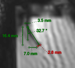
Within the department of Biomechanical Engineering at TU Delft we mainly focus on the development of novel medical instruments for minimally invasive therapy. The novel Steerable Needle Design to optimize LDR brachytherapy interventions is being conceived within CoBra Project framework by the PhD researcher Martijn de Vries. Steerable needles should allow for: better control of the needle track and end location during needle placement and allow for a large reach within the prostate for a more homogeneous dose. Needle steering is achieved using a compliant mechanism integrated in the needle tip, which can be actuated at the proximal end. Linear movements of the motors on the proximal end allows for steering at the distal end of the system. Preliminary tests under 3 T magnetic field show the feasibility of such design for application in-bore under live-MRI. The needle tip was steered in agar-agar phantom, actuating the needle in open-loop while tracking the needle tip under live MRI. During the test, the tip was bent approximately 7 mm and was trackable. The next steps are shielding of electronics for improved imaging and optimising the selection of imaging sequence.
OPTIMIZED NEEDLE TRAJECTORY
University of Portsmouth, Portsmouth, UK
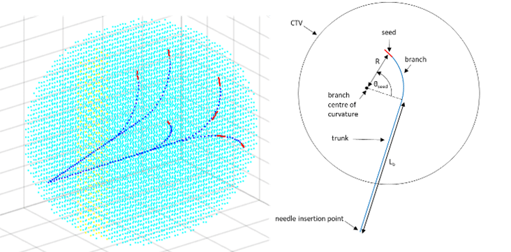
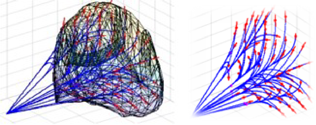
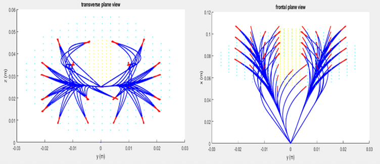
Based on the steerable needle developed by TU Delft, the task of the University of Portsmouth team was to develop a trajectory planning and optimisation algorithm for the path of this needle. The trajectory optimisation is intended to minimise tissue damage while avoiding organs at risk (neurovascular bundles, ureter). Hence it provides a more accurate and flexible method, which can also potentially both increase the scope of such procedure and reduce the side effects.
COBRA ROBOT INTEGRATION WITHIN THE MR COMPATIBLE AUTOMATED ROBOT GUIDE
University of Lille, Villeneuve d’Ascq, France
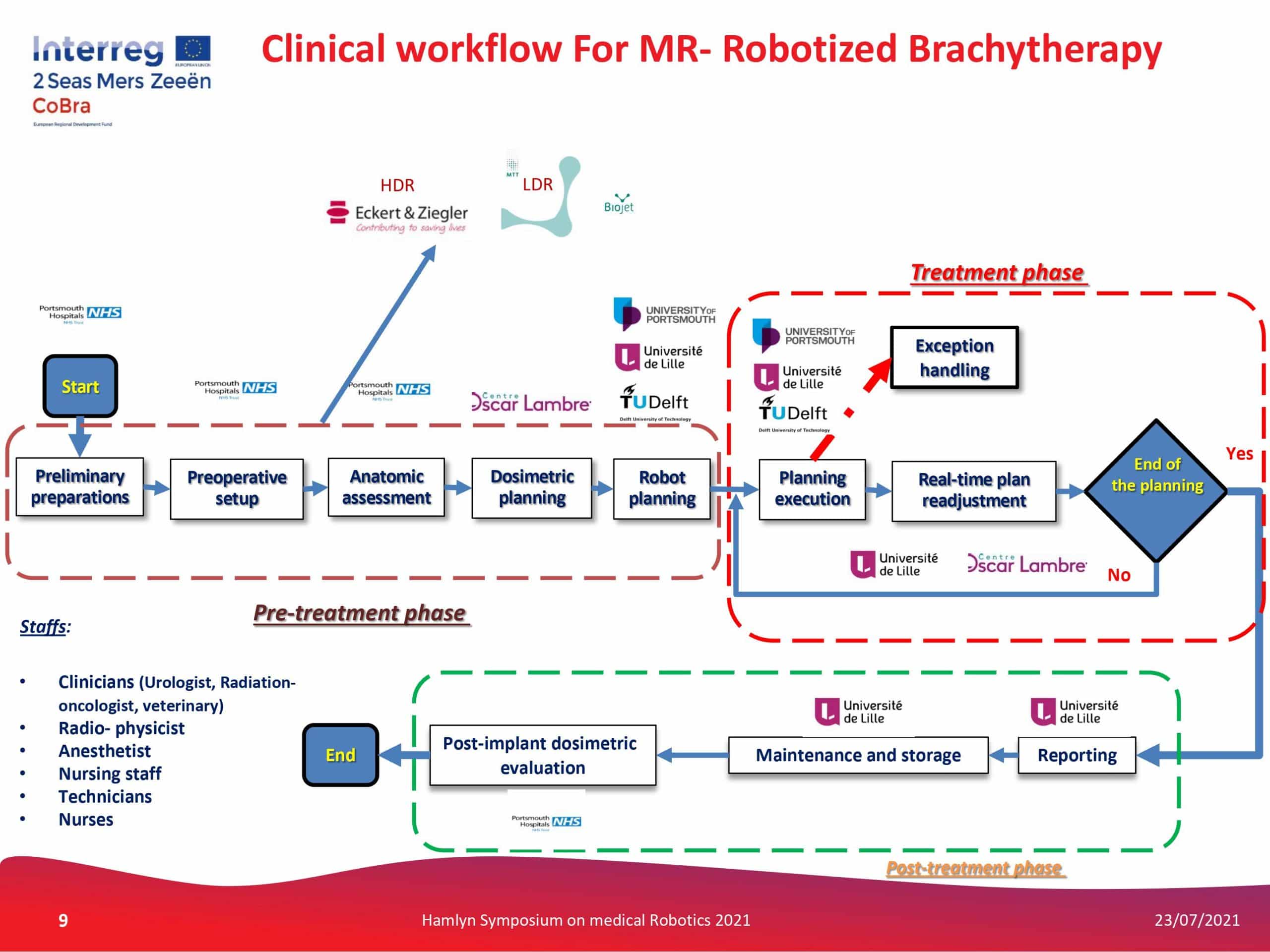
The integrated CoBra robotic solution is composed of the automated robot guide with 5 degrees of freedom, with interchangeable Biopsy Module or Brachytherapy Module with steerable needle and the seed loader developed in the University of Lille. The robot guide is composed of a parallel robot mechanism sized to cover a 3D workspace of prostate organ area accessible from lithotomy position of the patient. It can be controlled in closed loop jointly in position and velocity. This is due to the presence of encoders and absolute sensors for their joints and links operating in 3 Tesla MR fields.
The software basis is represented by the integration of the dose calculation and trajectory construction algorithms for treatment planning in real time with regards to MR imaging, the simulation platform coupled with bio-inspired phantom for tests and trainings.
The main advantages of the CoBra solution are its high-field MR-compatibility, real-time adaptation, precision, automation, and time-efficiency, all of which contribute to overall performance in diagnosis and treatment of localized cancers.
BIO-INSPIRED, SIMULATION-COUPLED, ACTIVE PROSTATE PHANTOM
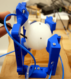
In the scope of the CoBra project, a new concept for an active, bio-inspired phantom (BIP) of the prostate was developed. The motivation behind the development of such an active-phantom is to mimic the real-organ behaviour during needle interventions for intra-operative treatment, e.g., brachytherapy. During the needle intervention, the prostate undergoes translational and rotational motions, and shows oedema due to the tissue damages. These factors result in lesion/target shift and cause inaccuracies in the dose-deposition. To achieve the desired dosimetry during robotised seed delivery, it is necessary to track the shift of lesions due to these factors (translation, change in orientation, and oedema). The novelty of the presented BIP is that it can mimic and sense both the prostate motions and oedema during needle interventions. It is coupled to the simulation framework SOFA through sensors inside the BIP in order to estimate the deformations and external forces. The BIP can be coupled with a robotic system to help in the study of automated needle insertions with adaptive control for precise dosimetry. The BIP is MRI-safe and can be used in-bore, allowing the accuracy validation of intra-operative robotic needle interventions prior to the clinical studies. The shown BIP proof-of-concept, which is in fact an anatomical soft robot, can potentially be extended towards further organs in future.
